
| Semipalatinsk nuclear test site (STS) (Kazakhstan) |
The Semipalatinsk Nuclear Test Site
(STS) also known as “Polygon” is located on northeast Kazakhstan being
attached to the Lrtysh river. The STS has been a primary venue for the
nuclear test site of the former Soviet Union. Since the first nuclear test
on September 1949, about 456 test explosions were performed until closure
of STS on August 29, 1991. All together approximately one million residents
of the villages near the test site were estimated to be exposed to radioactive
fallout, mostly from the fallout of atmospheric explosions performed until
1962. The explosions thereafter were underground explosions. Hydrogen fusion
explosion performed (-178m underground explosion with yield of 140 kt TNT
equivalent) on January 15, 1965 resulted in a large lake of about 400 m
diameter and 100 m depth (Chagan Lake; i.e., also known as “atomic lake”).
| [1] Preliminary study on chromosome aberrations, GPA- and TCR-mutation at STS (RBC, Kyoto University) |
Blood samples were collected from residents of Kurchatov, Akzhar and Sarzhal on May 16-19, 2000. Blood samples were immediately mixed with equal amount of “cold stimulating medium*” and kept in the cool (below 20 oC). The blood samples were brought by plane to RBC, Kyoto University, where lymphocyte cultures were established on May 26, 2000 for chromosome aberration analysis, and a part of the blood sample (still in the cold stimulating medium kept in the cold) was mailed to Radiation Effects Research Foundation, where GPA and TCR mutations were analysed by Dr. S. Kyoizumi and Dr. T. Seyama. The results are presented elsewhere.
Commentary
The cold stimulation method, which has been
introduced in IAEA Technical Reports in 2001 and 2011. The method has been
devised to keep the lymphocytes viable for a long time after blood sampling.
However, unfortunately the method has not been applied as originally recommended,
in particular, some reports use regular culture media, such as RPMI, and
attempt to keep nutritional condition and pH by adding L-glutamine and
HEPES. The original protocol recommends the use of Leibovitz’s L-15 medium. This culture
medium is the one specifically produced to culture in ambient atmosphere
without aeration of CO2 gas. The L-15 medium is composed of 10 times
amounts of amino acids, i.e., the amino acid-based buffered against
acidification by respiration of red blood cells, and high supply of nutrition
as well. The use of other commercially available culture media resulted in
earlier death of lymphocytes.
Sasaki, M. S. (1998): Pulling lymphocyte viability for long-term transportation of blood
samples. IAEA Symposium on Cytogenetic Analysis for Radiation Dose Assessment.
June 29-July 2, 1998. Budapest, Hungary.
IAEA (2001): Cytogenetic Analysis for Radiation
Dose Assessment: A Manual. IAEA Technical reports series No. 405. International
Atomic Energy Agency.
IAEA
(2011): Cytogenetic Dosimetry: Applications in Preparedness for and Response to
Radiation Emergencies. International Atomic Energy Agency.
| [2] Residents of Dolon (Testa et al. 2001) |
| Reference |
|
Testa, A., Stronati, L., Ranaldi, R., Spano, M., Steinhausler, F., Gastberger, M., Hubmer, A., Ptitkaya, L. and Akhmetov, M. (2001): Cytogenetic biomonitoring carried out in a village (Dolon) adjusent to the Semipalatinsk nuclear weapon test site. Radiat. Environ. Biophys., 40:125-129. |
| Study design |
|
Blood samples were collected from inhabitants of Dolon who have been living since 1949 (time of the first test explosion). The blood samples were immediately placed into polyform boxes and transferred by plane to the ENEA laboratory in Italy (with 36 hrs). Whole blood cultures were established, and chromosome preparations were stained with Giemsa. Control samples were collected from personnel of the Institute of Radiation Safety and Ecology of Kuruchatov. They were mainly involved in the administrative works and any occupational radiation exposure could be exclude. |
| Observations |
|
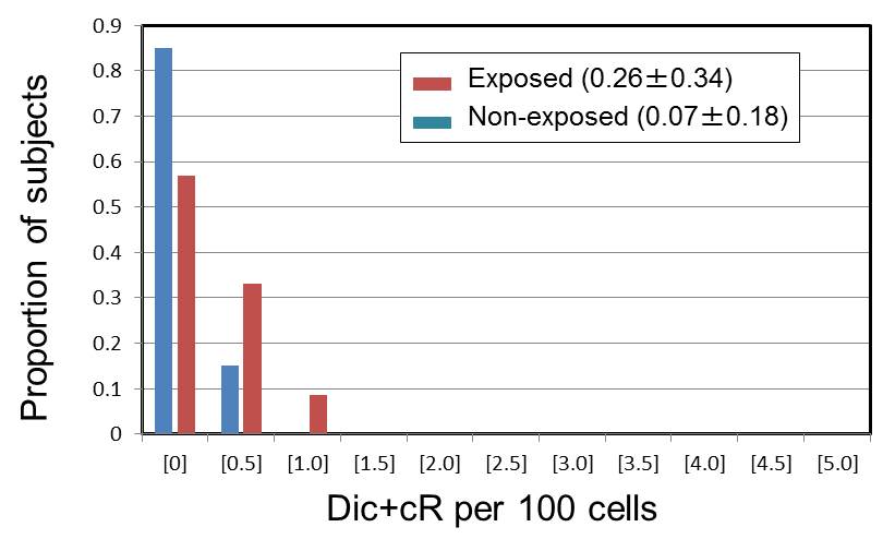 The distributions suggest difference between the exposed and non-exposed groups. |
||||||||||||||||||||||||||||||||||||||||||||||||||||||||||||||||||||||||||||||||||||||||||||||||||||||||||||||||||||||||||||||||||||||||||||||||||||||||||||||||||||||||||||||||||||||||||||||||||||||||||||||||||||||||||||||||||||||||||||||||||||||||||||||||||||||||||||||||||||||||||||||||||||||||||||||||||||||
| [3] Residents of areas near the STS (Tanaka et al. 2006) |
| Reference |
|
Tanaka, K., Iida, S., Takeichi, N., Chaizhunusova, N. J., Gusev, B. I., Aspalikov, K. N., Inaba, T. and Hoshi, M. (2006): Unstable-type chromosome aberrations in lymphocytes from individuals living near Semipalatinsk nuclear test site. J. Radiat. Res., 47(suppl):A159-A164. |
| Study design |
|
Blood samples were obtained from healthy
residents of differently contaminated areas near STS, Dolon, Sarjar, Kaynar,
Znamenka, Semipalatinsk and Socialistic, and remotely located uncontaminated
area, Kokpekty. The residents had been living in the areas between 1948
and 1965 when the atmospheric nuclear tests were performed. |
| Observations |
| [1] Unstable-type chromosome aberrations | [2] Micronuclei (MN) | ||||||||||||
| Villages | No. of | Age | No. of | No. of | Aerrations | No. of | No. of | No. of cells | Mean no. of | No. of MN | |||
| subjects | (mean±SD) | cells | abnormal cells | Dic* | cR | subjects | cells (mean) | with MN (mean) | MN/subject | per 1000 cells | |||
| Exposed | Dolon | 35 | 58.5±5.18 | 9,794 | 62 | 17 (9) | 8 | 53 | 5,018.5 | 46.8 | 50.8 | 9.36±3.46 | |
| Sarjar | 48 | 56.9±5.36 | 13,642 | 141 | 16 (6) | 7 | 44 | 4,098.3 | 38.9 | 42.2 | 9.9±3.62 | ||
| Kaynar | 33 | 56.9±4.84 | 11,650 | 40 | 14 (8) | 4 | 43 | 5,132.4 | 50.2 | 55.8 | 9.87±3.62 | ||
| Znamenka | 3 | 49.7±0.57 | 1,340 | 41 | 5 (0) | 4 | 10 | 5,112.1 | 35.9 | 35.9 | 7.1±3.0 | ||
| Semipalatinsk | 4 | 51.3±1.26 | 803 | 21 | 2 (0) | 2 | 42 | 4,734.2 | 61.8 | 61.8 | 12.3±3.94 | ||
| Socialistic | - | - | - | - | - | - | 20 | 4,348.4 | 31.8 | 34.1 | 7.3±3.08 | ||
| Controls | Kokpekty | 46 | 52.1±3.34 | 14,192 | 66 | 9 (6) | 2 | 21 | 5,034.5 | 36.5 | 38.5 | 7.25±2.14 | |
| *) Number in parentheses shows dicentrics with asssociated fragments. | |||||||||||||
| **) Higher aberration frequencies were found in some of the contaminated areas, but none was statistically significant as compared to the values in the control populations. | |||||||||||||
| [4] Residents of Dolon (Stephan et al. 2001) |
| Reference |
| Stephan, G., Pressl, S., Koshpessova. And Gusev, B. I. (2001): Analysis of FISH-painted chromosomes in individuals living near the Semipalatinsk nuclear test site. Radiat. Res., 155:796-800. |
| Study design |
| Blood samples were collected from 10 subjects
who were born before the first explosion in 1949 and lived continuously
in the village of Dolon. Dolon is highly polluted area among other villages.
hromosome translocations were analyzed by FISH painting method using whole chromosome painting probes for chromosomes 2, 4 and 8. The external dose to the inhabitants was estimated based on the physical measurements of dose rates along the track of radioactive clouds from explosion. Data derived from radiochemical analyses of soil, water, vegetation and food samples were used to estimate the internal dose. In Dolon, the effective external dose contributed about 48 % and the internal dose about 52 % of the total effective equivalent dose (B. J. Gusev, personal communication cited therein). |
|
Observations |
| Subject | Age* | Effective equivalen dose (Sv) | No. of | Chromosome aberrations** | Cells with | |||||||
| ID | (years) | External | Internal | Total | cells | tc | ti | dicc | dici | del | cr | complex aberrations |
| KD1 | 0.2 | 1.61 | 1.72 | 3.33 | 2,484 | 3 | 4 | 1 | ||||
| KD2 | 2.8 | 1.61 | 1.73 | 3.34 | 2,073 | 6 | 1 | 3 | 2 | |||
| KD3 | 2.0 | 1.61 | 1.73 | 3.34 | 2,172 | 13 | 3 | 2 | 2 | 1 | ||
| KD4 | 12.2 | 1.61 | 1.44 | 3.05 | 2,005 | 3 | 1 | 3a | ||||
| KD5 | 2.2 | 1.61 | 1.73 | 3.34 | 2,121 | 4 | 1 | 1 | ||||
| KD6 | 1.4 | 1.61 | 1.73 | 3.34 | 2,455 | 2 | 6b | 1 | 1 | 3 | ||
| KD7 | 2.9 | 1.61 | 1.73 | 3.34 | 2,192 | 2 | 1 | 1 | ||||
| KD8 | 4.3 | 1.61 | 1.73 | 3.34 | 2,555 | 2 | 2c | 1 | 1 | |||
| KD9 | 3.3 | 1.61 | 1.73 | 3.34 | 2,123 | 3 | 1 | 3 | ||||
| KD10 | 3.2 | 1.61 | 1.73 | 3.34 | 2,060 | 2 | 1 | |||||
| *) Age: age at the first explosion in 1949. | ||||||||||||
| **) t: translocation. dic: dicentrics. del: deletions. cr: centric rings. Subscripts (c) and (i) are "complete" and "incomplate" exchanges, respectively. | ||||||||||||
| a): One cell containing t(Ba)+t(Ab)+2t(Ab)+dic(AB); one cell with all labelled chromosome damaged. | ||||||||||||
| b) One translocation in combination withone dic and one t(bAA). | ||||||||||||
| c) One translocation in combination with one ring. | ||||||||||||
| [5] Residents of districts near the STS (Salomaa et al. 2002) |
| Reference |
|
Salomaa, S., Lindholm, C., Tankimanova, M. K., Zh. Mamyrbaeva, Z., Koivistoinen, A., Hulten, M., Mustonen, R., Dubrova, Y. E. and Bersimbaev, R. I. (2002): Stable chromosome aberrations in the lymphocytes of a population living in the vicinity of the Semipalatinsk nuclear rest site. Radiation Res., 158:591-596. |
| Study design |
|
The frequencies of stable-type chromosome aberrations have been studied by FISH chromosome painting technology in the inhabitants of villages near the Semipalatinsk nuclear test site (STS). |
| Conclusion The translocation frequencies were not significantly different among P0, P1 generations living in contaminated district and controls living in non-contaminated district. The observations could not substantiate the previously reported does of the order of 1-4.5 Gy to P0 generation in the contaminated area. Moreover, the age-dependent increase of translocation frequencies was not diferent between the exposed and control populations. |
| Generations | FISH analysis | Village exposed | Controls | |||||||
| Bodene | Chagan | Cheremushki | Dolon | Kanonerka | Karamyrza | Mostik | All combined | |||
| P0 | No. of subjects | 8 | 1 | 2 | 10 | 8 | 1 | 1 | 31 | 19 |
| No. of cells analysed | 15,800 | 2,000 | 3,860 | 19,243 | 13,752 | 1,105 | 753 | 56,513 | 40,743 | |
| Two-way translocations* | 8.7±1.3 | 5.7±2.8 | 7.7±2.3 | 9.5±1.2 | 4.8±1.0 | 5.2±3.7 | 3.8±3.8 | 7.7±0.6 | 8.1±0.8 | |
| All translocations* | 10.8±1.4 | 8.6±3.5 | 8.9±2.6 | 11.9±1.5 | 8.1±1.3 | 5.2±3.7 | 7.6±5.4 | 10.0±0.7 | 10.2±0.8 | |
| P1 | No. of subjects | 12 | - | 3 | 3 | 10 | - | - | 28 | 21 |
| No. of cells analysed | 24,276 | - | 7,231 | 6,000 | 18,029 | - | - | 55,536 | 43,999 | |
| Two-way translocations* | 1.5±0.4 | - | 4.7±1.4 | 2.4±1.1 | 4.0±0.8 | - | - | 2.8±0.4 | 5.8±0.6 | |
| All translocations* | 2.0±0.4 | - | 7.9±1.8 | 2.4±1.1 | 5.7±0.9 | - | - | 4.0±0.5 | 5.8±0.6 | |
| *) Translocations per 1000 cell equivalent ± SE. Two-way translocation: t(Ab)+t(Ba). Other translocation: t(Ab) or t(Ba). | ||||||||||
| [6] Residents of Dolon and Chekoman (Chaizhunusova et al. 2006) |
| Reference |
|
Chaizhunusova, N., Yang, T. C., Land, C., Luckyanov, N., Wu, H., Apsalikov, K. N. and Madieva, M. (2006): Biodosimetry study in Dolon and Chekoman villages in the vicinity of Semipalatinsk nuclear test site. J. Radiat. Res., 47 (suppl):A165-A169. |
| Study design |
|
A total of 15 women were selected from Dolon
and 15 women from Chekoman districts. They were all born before the first
nuclear test in August 1949. Chromosome aberrations in blood lymphocytes
were studied by FISH painting technique. |
| Observations |
| [1] Dolon | [2] Chekoman | |||||||
| Subject | Cells | Aberrations | Subject | Cells | Aberrations | |||
| ID | scored | Number | Genome equivalent | ID | scored | Number | Genome equivalent | |
| D1 | 964 | 8 | 26.4 | C1 | 851 | 1 | 3.3 | |
| D2 | 221 | 0 | 0 | C2 | 2011 | 5 | 16.5 | |
| D3 | 3348 | 11 | 36.3 | C3 | 283 | 0 | 0 | |
| D4 | 3562 | 9 | 29.7 | C4 | 934 | 1 | 3.3 | |
| D5 | 1105 | 5 | 16.5 | C5 | 712 | 0 | 12.4 | |
| D6 | 132 | 1 | 3.3 | |||||
| D7 | 181 | 0 | 0 | |||||
| D8 | 164 | 1 | 3.3 | |||||
| D9 | 58 | 1 | 3.3 | |||||
| D10 | 2723 | 19 | 62.7 | |||||