| Dose-response parameters and microdosimetric quantity [1] |
| Charged particles and microdose |
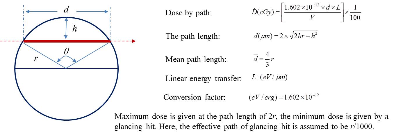
Biologically important dose is given by charged particle (also Compton
or photo electrons in the case of photons, and recoil protons or other
charged particles in the case of neutrons). When the average dose incident
to the cell nucleus is decreased, the dose to the cell nucleus does not
proportionally decrease at low doses, where the dose to the hit nucleus
is defined only by the quality of the charged particle (LET), and hence
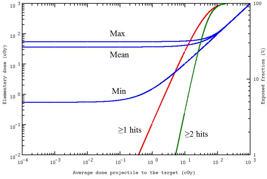
only the number of the hit cell nuclei decreases. This minimum dose given
by traversal of single charged particle is called “elementary dose” or
microdose”. The followings are example of dose calculation for a charged
particle (5 MeV alpha particle with LET=132.24 keV/μm passing through a
lymphocyte nucleus with 5 μm diameter.
| Electrons and beta-particles |
| Electrons and β-particles |
|
|
|
|
|
|
|
|
|
|
|
|
|
|
|
|
|
| Radiation |
Mean energy |
Mean LET (keV/μm) |
Authors |
Dose range |
Data |
Linear term (×10-4/cell/cGy) |
Quadratic term (×10-6/cell/cGy2) |
Microdose |
σ=Dics/cell/path |
| (MeV) |
LT |
LT(cutoff) |
LD |
LD(cutoff) |
(cGy) |
points |
α |
± |
(S.E.) |
β |
± |
(S.E.) |
(cGy) |
σ±SE |
| Electrons(100rad/min) |
15 |
0.214 |
0.106 |
0.222 |
0.117 |
Purrott et al. 1977 |
44-511 |
6 |
0.400 |
± |
1.094 |
6.537 |
± |
0.425 |
0.17 |
6.800E-06 |
± |
1.860E-05 |
| Electrons(pulsed) |
15 |
0.214 |
0.106 |
0.222 |
0.117 |
Purrott et al. 1977 |
53-392.2 |
7 |
0.200 |
± |
1.835 |
8.093 |
± |
1.141 |
0.17 |
3.400E-06 |
± |
3.120E-05 |
| Electrons |
3 |
0.204 |
0.113 |
0.233 |
0.135 |
Bauchinger et al. 1974 |
128-432** |
9 |
1.000 |
± |
2.395 |
3.488 |
± |
0.485 |
0.17 |
1.700E-05 |
± |
4.072E-05 |
| 90Sr-90Y beta |
0.6013 |
0.580 |
0.126 |
0.484 |
0.126 |
Vulpis et al. 1986 |
13.8-276 |
7 |
8.490 |
± |
1.566 |
8.698 |
± |
0.722 |
0.22 |
1.868E-04 |
± |
3.445E-05 |
| 90Y beta |
2.281 |
0.413 |
|
|
|
Schmid et al. 2006 |
0-217.6 |
7 |
2.290 |
± |
0.280 |
0 |
± |
0 |
0.34 |
7.786E-05 |
± |
9.520E-06 |
| 3H beta (80% water) |
0.0057 |
5.594 |
2.977 |
6.717 |
3.025 |
Vulpis 1984 |
25-420 |
8 |
12.935 |
± |
1.634 |
5.731 |
± |
0.611 |
4.56 |
5.898E-03 |
± |
7.451E-04 |
| 3H beta (82% water) |
0.0057 |
5.594 |
2.977 |
6.717 |
3.025 |
Prosser et al. 1983 |
10.2-304.5 |
10 |
5.652 |
± |
0.403 |
1.929 |
± |
0.172 |
4.56 |
2.577E-03 |
± |
1.838E-04 |
| 3H beta (70% water) |
0.0057 |
5.594 |
2.977 |
6.717 |
3.025 |
Tanaka et al. 1994 |
0-210 |
6 |
20.639 |
± |
2.539 |
8.544 |
± |
1.504 |
4.56 |
9.411E-03 |
± |
1.158E-03 |
| 3H beta (80% water)* |
0.0057 |
5.594 |
2.977 |
6.717 |
3.025 |
Bocian et al. 1977 |
0-255 |
5 |
8.492 |
± |
1.048 |
9.353 |
± |
0.851 |
4.56 |
3.872E-03 |
± |
4.779E-04 |
| *) Including centric rings. |
|
|
|
|
|
|
|
|
|
|
|
|
|
|
|
|
|
|
| **) Because there are no low-dose points, the data were fitted to Y=C+αD+βD2 |
|
|
|
|
References
Bauchinger, M., Schmid, E. and Rimpl, G. (1974): Interaction distance of primary lesions in the formation of dicentric chromosomes after irradiation of human lymphocytes with 3-MeV electrons in vitro. Mutation
Res., 25:83-87.
Bocian, E., Ziemba-Zak, B., Rosiek, O. and
Sablinski, J. (1977): Chromosome aberrations in human lymphocytes exposed
to tritiated water in vitro. Top. Radiat. Res. Quat., 12:168-181.
Prosser, J. S., Lloyd, D. C., Edwards, A.
A. and Stather, J. W. (1983): The induction of chromosome aberrations in
human lymphocytes by exposure to tritiated water in vitro.
Radiat. Prot. Dosimet., 4:21-26.
Purrott, R. J., Reder, E. J. and Lovell, S. (1977): Chromosome aberration yields induced in human lymphocytes by 15 MeV electrons given at a conventional dose-rate and in microsecond pulses. Int. J. Radiat. Biol., 31: 251-256.
Schmid, E., Rimpl, G. and Bauchinger, M.
(1974): Dose-response relation of chromosome aberrations in human lymphocytes
after in
vitro irradiation with 3-MeV electrons. Radiat. Res., 57:228-238.
Tanaka, K.,
Sawada, S. and Kamada, N. (1994): Relative biological effectiveness and dose
rate effect of tritiated water on chromosomes in human lymphocytes and bone
marrow cells. Mutation Res., 323:53-61.
Vulpis, N. (1984): The induction of chromosome
aberrations in human lymphocytes by in vitro irradiation with β particles
from tritiated water. Radiat. Res., 97:511-518.
Vulpis, N. and Scarpa, G. (1986): Induction
of chromosome aberrations by 90Sr β-particles
in cultured human lymphocytes. Mutation Res., 163:277-283.
| Protons |
|
|
|
|
|
|
|
|
|
|
|
|
|
|
|
| Radiation |
Energy |
Mean LET |
Authors |
Dose range |
Data |
Linear term (×10-4/cell/cGy) |
Quadratic term (×10-6/cell/cGy2) |
Microdose |
σ=Dics/cell/path. |
| (MeV) |
LT (keV/μm) |
(cGy) |
points |
α |
± |
(S.E.) |
β |
± |
(S.E.) |
ε (cGy) |
σ±SE |
| Protons |
8.7 |
5.17 |
Edwards et al. 1986 |
6.2-279 |
9 |
6.838 |
± |
0.777 |
4.362 |
± |
0.342 |
4.22 |
2.886E-03 |
± |
3.279E-04 |
| Protons |
40 |
1.47 |
Sasaki 2009 |
0-300 |
4 |
1.086 |
± |
1.621 |
4.362 |
± |
0.700 |
1.20 |
1.303E-04 |
± |
1.945E-04 |
| Protons |
60 |
1.07 |
Manti et al. 2005 |
0-500 |
6 |
0.660 |
± |
0.360 |
1.480 |
± |
0.090 |
0.87 |
5.742E-05 |
± |
3.132E-05 |
| Protons |
4.9 |
7.9 |
Takatsuji et al. 1983 |
16-310 |
12 |
28.018 |
± |
11.021 |
14.568 |
± |
5.688 |
6.45 |
1.807E-02 |
± |
7.109E-03 |
| Protons |
22.6 |
4.21 |
Joksic et al. 2000 |
0-300 |
5 |
18.150 |
± |
2.040 |
0.705 |
± |
0.729 |
3.43 |
6.225E-03 |
± |
6.997E-04 |
| Protons |
50 |
1.23 |
Todorov 1972, 1975 |
0-250 |
5 |
0.877 |
± |
1.466 |
4.698 |
± |
0.712 |
1.00 |
8.770E-05 |
± |
1.466E-04 |
| Protons |
16.5 |
2.14 |
Schmid et al. (D1) 1997,Ld=3.5 |
0-132 |
6 |
4.580 |
± |
0.271 |
1.805 |
± |
0.239 |
1.75 |
8.015E-04 |
± |
4.743E-05 |
| Protons |
16.5 |
3.15 |
Schmid et al. (D2) 1997, Ld=5.3 |
0-198 |
6 |
6.500 |
± |
1.000 |
1.400 |
± |
0.300 |
2.57 |
1.671E-03 |
± |
2.570E-04 |
| Protons |
16.5 |
11.3 |
Schmid et al. (D3) 1997, Ld=19 |
0-145 |
6 |
32.300 |
± |
1.300 |
0 |
± |
0 |
9.22 |
2.978E-02 |
± |
1.199E-03 |
| Protons |
16.5 |
1.8 |
Rimpl et al. 1990, Ld=3 |
14-287 |
8 |
4.400 |
± |
0.700 |
1.950 |
± |
0.300 |
1.47 |
6.468E-04 |
± |
1.029E-04 |
| Protons |
31 |
1.83 |
Bettega et al. 1981 |
0-200 |
5 |
6.260 |
± |
0.484 |
1.815 |
± |
0.286 |
1.49 |
9.327E-04 |
± |
7.212E-05 |
| Protons |
12 |
3.99 |
Bettega et al. 1981 |
0-200 |
5 |
9.189 |
± |
1.395 |
3.501 |
± |
0.835 |
3.26 |
2.996E-03 |
± |
4.548E-04 |
| Protons |
8 |
5.82 |
Bettega et al. 1981 |
0-200 |
5 |
24.950 |
± |
5.770 |
2.797 |
± |
3.582 |
4.75 |
1.185E-02 |
± |
2.741E-03 |
| Protons |
70 |
1 |
Matsubara et al. 1990 (5mm) |
10-300 |
6 |
2.195 |
± |
1.842 |
4.659 |
± |
0.550 |
0.82 |
1.800E-04 |
± |
1.510E-04 |
| Protons |
70 |
1.15 |
Matsubara et al. 1990(15mm) |
10-300 |
6 |
7.508 |
± |
2.184 |
4.073 |
± |
0.927 |
0.94 |
7.058E-04 |
± |
2.053E-04 |
| Protons |
70 |
1.42 |
Matsubara et al. 1990 (25mm) |
10-300 |
6 |
9.602 |
± |
0.691 |
4.110 |
± |
0.267 |
1.16 |
1.114E-03 |
± |
8.016E-05 |
| Protons |
1000 |
0.31 |
Repina et al. 2007 |
5-200 |
|
1.200 |
± |
0.200 |
0 |
± |
0 |
0.25 |
3.000E-05 |
± |
5.000E-06 |
| Protons |
7.4 |
5.84 |
Bocian 1973 (Lloyd and Edwards 1983) |
50-400 |
|
15.100 |
± |
5.640 |
0.530 |
± |
0 |
4.76 |
7.188E-03 |
± |
2.685E-03 |
| Protons |
0.88 |
28 |
Mognato et al. 2003 |
0-200 |
6 |
61.800 |
± |
8.900 |
0 |
± |
0 |
22.84 |
1.412E-01 |
± |
2.033E-02 |
References
Bettega, D., Dubini, S., Conti, A. M. F,
Pelucchi, T. and Lombardi, L. T. (1981): Chromosome aberrations induced
by protons up to 31 MeV in cultured human cells. Radiat. Environ. Biophys.,
19: 91-100.
Edwards, A. A., Lloyd, D. C., Prosser, J. S., Finnon, P. and Moquest, J. E. (1986): Chromosome aberrations induced in human lymphocytes by 8.7 MeV protons and 23.5 MeV helium-3 ions. Int. J. Radiat. Biol., 50: 137-145.
Joksie, G.,
Pajovic, S. B., Stankovic, M., Pejic, S., Kasapovic, J., Guttone, G., Calonghi,N.,
Masotti, L. and Kanazir, D. T. (2000): Chromosome aberrations, micronuclei, and
sctivity of superoxide dismutases in human lymphocytes after irradiation in
vitro. Cell. Mol. Life Sci., 57:842-850.
Matsubara, S., Ohara, H., Hiraoka, T., Koike,
S., Ando, K., Yamaguchi, H., Kuwabara, Y., Hoshina, M. and Suzuki, S. (1990):
Chromosome aberration frequencies produced by a 70-MeV proton beam. Radiation
Research, 123, 182-191.
Manti, L., Durante, M., Cirrone, G. A., Grossi, G., Lattuada, M., Sabini, M. G., Scampoli,P., Valastro, L. and Gialanella, G. (2005): Modelled microgravity does not modify the yield of chromosome aberrations induced by high-energy protons in human lymphocytes. Int. J. Radiat. Biol., 81:147-155.
Mognato, M., Bortoletto, E., Ferraro, P.,
Baggio, L., Cherubini, R., Canova, S., Russo, A. and Celotti, L. (2003):
Genetic ddamage induced by in vitro irradiation of human G0 lymphocytes with
low-energy protons (23 keV/microm): HPRT mutations and chromosomeaberrations.
Radiat. Res., 160:52-60.
Repina, L. A., Abrosimova, A. N., Timoshenko,
G. N. and Issinskii, I. B. (2007): Cytogenetic effects in human lymphocytes
in vitro after irradiation by protons with 1-GeV energy. Aviakosm. Ekolog.
Med., 41:29-32 (Russian).
Rimple, G. R., Schmid, E., Braselmann, H. and Bauchinger, M. (1990): Chromosome aberrations induced in human lymphocytes by 16.5 MeV protons. International Journal of Radiation Biology, 58, 999-1007.
Sasaki, M. S. (2009): Advances in the biophysical
and molecular basis of radiation cytogenetics. Int. J. Radiat. Biol., 85:26-47.
Schmid, E., Roos, H., Rimple, G. and Bauchinger,
M. (1997): Chromosome aberration frequencies in human lymphocytes irradiated
in a multi-layer array by protons with different LET. International Journal
of Radiation Biology, 72, 661-665.
Takatsuji, T., Takekoshi, H. and Sasaki, M. S. (1983): Induction of chromosome aberrations by 4.9 MeV protons in human lymphocytes. International Journal of Radiation Biology, 44, 553-562.
Todorov, S. L., Grigor’ev, Y. G., Rizhov,
N. I., Ivanov, B. A., Malyutina, T. S. and Mileva, M. S. (1972) Dose-response
relationship for chromosome aberrations induced by X-rays or 50 MeV protons
in human peripheral lymphocytes. Mutation Research, 15, 215-220.
| Alpha-particles |
|
|
|
|
|
|
|
|
|
|
|
|
|
|
|
| Radiation |
Mean |
Mean LET |
Authors |
Dose range |
Data |
Linear term (×10-4/cell/cGy) |
Quadratic term (×10-6/cell/cGy2) |
Microdose |
σ=Dics/cell/path. |
| energy (MeV) |
LT (keV/μm) |
(cGy) |
points |
α |
± |
(S.E.) |
β |
± |
(S.E.) |
(cGy) |
σ |
± |
SE |
| 23 MeV α |
23 |
28.03 |
Takatsuji et al. 1984 |
25.6-299 |
7 |
74.180 |
± |
6.190 |
2.234 |
± |
5.544 |
22.87 |
1.696E-01 |
± |
1.416E-02 |
| 18 MeV α |
18 |
34.01 |
Sasaki et al. 1998 |
27-270 |
5 |
70.230 |
± |
6.510 |
4.853 |
± |
2.913 |
27.75 |
1.949E-01 |
± |
1.807E-02 |
| 8.4 MeV α |
8.4 |
61.8 |
Sasaki et al. 1998 |
30-150 |
4 |
96.390 |
± |
10.570 |
4.183 |
± |
7.379 |
50.42 |
4.860E-01 |
± |
5.329E-02 |
| 3.85 MeV α |
3.85 |
109 |
Durante et al. 1995 |
0-200 |
7 |
44.080 |
± |
7.920 |
0 |
± |
4.088 |
88.93 |
3.920E-01 |
± |
7.043E-02 |
| Helium-3 |
23.5 |
21 |
Edwards et al. 1986 |
4.7-282 |
8 |
40.500 |
± |
3.200 |
0 |
± |
1.800 |
17.13 |
6.938E-02 |
± |
5.482E-03 |
| 241Am α |
5.49 |
125.9 |
DuFrain et al. 1979* |
0.85-6.84 |
8 |
95.135 |
± |
4.170 |
0 |
± |
0 |
102.72 |
9.772E-01 |
± |
4.283E-02 |
| 241Am α |
2.7 |
169.5 |
Schmid and Roos 2009 |
|
|
27.700 |
± |
2.000 |
0 |
± |
0 |
122.38 |
3.390E-01 |
± |
2.448E-02 |
| 241Am α |
2.7 |
150 |
Mestres et al. 2004 |
20-100 |
4 |
36.780 |
± |
3.210 |
1.689 |
± |
3.853 |
122.38 |
4.501E-01 |
± |
3.928E-02 |
| 241 Am α |
2.7 |
150 |
Schmid et al. 1996 |
0-100 |
9 |
26.910 |
± |
2.630 |
1.639 |
± |
3.455 |
122.38 |
3.293E-01 |
± |
3.219E-02 |
| 241 Am α |
2.7 |
150 |
Baraquinero et al. 2004 |
0-100 |
9 |
27.510 |
± |
3.960 |
1.879 |
± |
4.661 |
122.38 |
3.367E-01 |
± |
4.846E-02 |
| 241 Am α (PCC) |
3.45 |
127.15 |
Greinert et al. 1999 |
100-300 |
3 |
61.140 |
± |
1.758 |
0 |
± |
0 |
103.74 |
6.343E-01 |
± |
1.824E-02 |
| 225Ac/213Bi α |
4.95 |
138.568 |
Tawn et al. 2007, 2009 |
0-50 |
7 |
33.600 |
± |
0.470 |
0 |
± |
0 |
113.06 |
3.799E-01 |
± |
5.314E-03 |
| 238Pu α |
(3.5) |
155 |
Purrott et al. 1980 |
6.5-160 |
6 |
31.680 |
± |
2.570 |
4.696 |
± |
1.838 |
126.46 |
4.006E-01 |
± |
3.250E-02 |
| 239Pu α |
3.5 |
116 |
Cornforth et al. 2002 |
0-220 |
5 |
55.650 |
± |
5.590 |
0 |
± |
2.859 |
94.64 |
5.267E-01 |
± |
5.290E-02 |
| 242Cm α |
4.9 |
140 |
Edwards et al. |
10.6-282 |
9 |
28.410 |
± |
3.810 |
0 |
± |
1.591 |
114.22 |
3.245E-01 |
± |
4.352E-02 |
| *) The dose was re-evaluated by A. A. Edwards to be at least 3 times higher than that described (Edwards, A. A., Int. J. Radiat. Biol., 38:83-91, 1980. |
|
Commentary: When the dose is adjusted by lymphocyte survival (D0=3Gy), the dicentric response could be Y=(30.405±1.332)×10-4/cGy |
References
Barquinero, J. F., Stephan, G. and Schmid,
E. (2004): Effect of americium-241 α-particles on the dose-response of
chromosome aberrations in human lymphocytes analysed by fluorescence in situ hybridization.
Int. J. Radiat. Biol., 80:155-164.
Cornforth, M. N., Bailey, S. M. and Goodwin, E. H. (2002): Dose responses for chromosome aberrations produced in noncycling primary human fibroblast by alpha particles, and by γ rays delivered at sublimiting low dose rates. Radiat. Res., 158:43-53.
DuFrain, R. J.,
Littlefield, L. G., Joiner, E. E. and Frome, E. L. (1979): Human cytogenetic
dosimetry: a dose-response relationship for alpha particle radiation from 241Am.
Health Phys., 37:279-289.
Durante, M., Grossi, G. F., Gialanella, G.,
Pugliese, M., Nappo, M. and Yang, T. C. (1995): Effects of α-particles
on survival and chromosomal aberrations in human mammary epithelial cells.
Radiat. Environ. Biophys., 34: 195-204.
Edwards, A. A., Purrott, R. J., Prosser, J. S. and Lloyd, D. C. (1980): The induction of chromosome aberrations in human lymphocytes by alpha-radiation. Int. J. Radiat. Biol., 38:83-91.
Edwards, A. A.,
Lloyd, D. C., Prosser, J. S., Finnon, P. and Moquet, J. E. (1986): Chromosome
aberrations induced in human lymphocytes by 8.7 MeV and 23.5 MeV herium-3 ions.
Int. J. Radiat. Biol., 50:137-145.
Greinert, R., Thieke, C., Detzier, E., Boguhn,
O., Frankenberg, D. and Harder, D. (1999): Chromosome aberrations induced
in human lymphocytes by 3.45 MeV α particles analyzed by premature chromosome
condensation. Radiat. Res., 152:412-420.
Mestres, M., Caballin, M. R., Schmid, E., Stephan, G., Sachs, R., Barrios, L. and Barquinero, J. E. (2004): Analysis of α-particle induced chromosome aberrations in human lymphocytes, using pan-centromeric and pan-telomeric probes. Int. J. Radiat. Biol., 80:737-744.
Purrott, R. J., Edwards, A. A., Lloyd, D.
C. and Stather, J. W. (1980): The induction of chromosome aberrations in
human lymphocytes by in vitro irradiation with α-particles from plutonium-239.
International Journal of Radiation Biology, 38, 277-284.
Sasaki, M. S., Takatsuji, T. and Ejima, Y.
(1998): The F
value cannot be ruled out as a chromosomal fingerprint of radiation quality.
Radiat. Res., 150:253-258.
Sasaki, M. S.
(2009): Advances in the biophysical and molecular basis of radiation
cytogenetics. Int. J. Radiat. Biol., 85:26-47.
Schmid, E.,
Hieber, L., Heinzmann, U., Roos, H. and Kellerer, A. M. (1996): Analysis of
chromosome aberrations in human peripheral lymphocytes induced by in vitro α-particle
irradiation. Radiat. Environ. Biophys., 35: 179-184.
Schmid, E. and
Roos, H. (2009): Influence of the bystander phenomenon on the chromosome
aberration pattern in human lymphocytes induced by in vitro α-particle
exposure. Radiat. Environ. Biophys., 48:181-187.
Tawn, E. J. and Thierens, H. (2009): Dose response relationships for chromosome aberrations induced by low doses of alpha-particle radiation. Radiat. Prot. Dosimet., 135:268-271.
Takatsuji, T. and Sasaki, M. S. (1984): Dose-effect
relationship of chromosome aberrations induced by 23 MeV alpha particles
in human lymphocytes. Int. J. Radiat. Biol., 45: 237-243.
| Accelerated ions (HZE) |
|
|
|
|
|
|
|
|
|
|
|
|
|
| Radiation |
Mean energy |
Mean LET |
Authors |
Dose range |
Data |
Linear term (×10-4/cell/cGy) |
Quadratic term (×10-6/cell/cGy2) |
Microdose |
σ=Dics/cell/path |
| (MeV) |
LT (keV/μm) |
(cGy) |
points |
α |
± |
(S.E.) |
β |
± |
(S.E.) |
ε (cGy) |
σ |
± |
SE |
| Helium-4 |
20.5 |
31.4 |
Di Giorgio et al. 2004 |
0-302.9 |
9 |
37.3 |
± |
1.8 |
0 |
± |
0 |
25.62 |
9.556E+02 |
± |
4.612E+01 |
| Oxygen-16 |
1480 |
52 |
Di Giorgio et al. 2004 |
0-160 |
10 |
56.6 |
± |
4.5 |
0 |
± |
0 |
42.43 |
2.402E+03 |
± |
1.909E+02 |
| Carbon-12 |
425 |
61 |
Di Giorgio et al. 2004 |
0-147 |
9 |
67.4 |
± |
5.3 |
0 |
± |
0 |
49.77 |
3.354E+03 |
± |
2.638E+02 |
| Oxygen-16 |
996 |
69 |
Di Giorgio et al. 2004 |
0-222 |
9 |
38.5 |
± |
2.9 |
0 |
± |
0 |
56.3 |
2.168E+03 |
± |
1.633E+02 |
| Neon-20 |
212 |
460 |
Edwards et al. 1994 |
|
|
0.41 |
± |
0.015 |
0 |
± |
0 |
375.31 |
1.539E+02 |
± |
5.630E+00 |
References
Di Giorgio, M., Edwards, A. A., Moquest,
J. E., Finnon, P., Hone, P. A., Lloyd, D. C., Kreiner, A. J., Schuff, J.
A., Taja, M. R., Vallerga, M. B., López, F. O., Burlón, A., Debray, M.
E. and Valda, A. (2004): Chromosome aberrations induced in human lymphocytes
by heavy charged particles in track segment mode. Radiat. Protect. Dosimet.,
108: 47-53.
Edwards, A. A., Finnon, P., Moquet, J. E., Lloyd, D. C., Darroudi, F. and Natarajan, A. T. (1994): The effectiveness of high energy neon ions in producing chromosomal aberrations in human lymphocytes. Radiat. Protect. Dosimet., 52:299-203.