| 137Cs Accident, Estonia (1994) |
| Accident scenario |
| Recently, Estonia has become a bridge for
re-selling scrap metal from countries of the former Soviet Union to the
West. The discovery of radiation sources or radioactive pieces of metal,
especially in the scrap metal, is presently occurring more frcqucntly in
this country. Sixteen 137-Cs sources have bccn stolen from difrercnt Estonian
plants during the 1992-1994 period, and eleven unknown radiation sources
have been found in Estonia in 1993 and 1994. The most dramatic accident
occurred in the village Kiisa near Tauinn in l994, whcrc a high-activity
137-Cs source was discovered in the kitchen of one of the dwelling houses. On 21 October l994, three brothers broke into the Tammiku repository of radioactive wastes, some 20 km from Tallinn, with the aim to steal some metal and then sell it. During this ‘operation’, a radiation source fell out from one metal block. It had an appearance of a small metal cylinder, about 1.5 cm in diameter and 3 cm long. One of the brothers, R.H., took the cylinder and placed it into the pocket of his jacket. The next morning he came home to the village Kiisa (about 30 km from Tallinn) and put the jacket, presumably with the source inside, onto the coat hanger in the entrance hall. Not feeling well, he went to bed, and, according to one version, did not remove his jacket during this time. He was hospitalized on 25 October and died on 2 November due to acute radiation sickness. Nevertheless, the diagnosis was kidney failure, and apparently no one knew about the radiation source. Besides R.H., three members of his family lived in the house. His wife P.K., a 13 year old stepson R.T. and the boy's grandmother A.S. On 9 November, R.T. was repairing his bicycle. According to an official version, he found the source in a pocket of the jacket, put it into the drawer with tools and then returned the drawer to the kitchen. On l7 November, the boy was hospitalized with severe burns to his hands. These were recognized as radiation burns, and the police were notified. On 18 November the staff of the Rescue Board found the source in the drawer in the kitchen. The source has since been identified as 137-Cs from a sterilization source assembly. During the rescue operation, an exposure rate of 20 R/h was measured. |
| Early clinical findings |
| The two surviving brothers (subjects 1 and 2) who visited the waste depository were exposed on 21 October. According to anamnestic data, the exposure most probably lasted for about 7h, first at the depository and then during several hours they spent with the brother that was carrying the source in his pocket. In addition to whole-body exposure, both brothers also had burns on their hands, suggesting that they must have touched the source directly. The three residents (subjects 3-5) in the house where the source was located were exposed to whole-body radiation for 4 weeks from 21October to 17 November. A dose-rate >1 Gy/h was measured in the kitchen by the Rescue Board at a distance of about 1 m from the source. During most of the time spent indoors, however, the residents were exposed to a relatively low dose-rate ranging from several to tens mGy/h. Subject 3, a 13-year-old boy, had severe burns on his hands as a consequence of localized exposure while hand-ling the source. Three friends and neighbours (subjects 6-8) visited the house on some occasions during the 4 weeks; exact dates and durations of stay are not known, but subject 6, a 12-year-old boy, was present for several hours when subject 3 was repairing his bike. The other people examined (medical and rescue personnel) had been exposed during shorter visits to the house, the medical personnel (subjects 9-11) during 23-25 October and the rescue personnel (subjects 12-18) on 18 November. One member of the rescue team (subject 18) had touched the source for several seconds as he moved it to a lead container. When the source was discovered, eight persons (subjects 1-8) were hospitalized for observation. On admission on 22 November, thrombocytopaenia, 54 x 10(9)/l, subject 1 had which increased to normal values was 3.4 x 10(9)/l in 2 weeks. His leukocyte count and the absolute was 1.2 x 10(9)/1. While the lymphocyte count 1eukocyte count soon normalized, minimum of the lymphocyte count fell to a o.85 x 10(9)/l on 7 December. These values point to a mild degree of radiation sickness. No clinical symptoms were noted in addition to mild radiation burns on the hands. Subject 2 had a leukocyte count of 3.4 x 10(9)/l on admission (22 November); the other blood values were normal. This may be interpreted as a borderline sign of bone marrow depression. He also had severe radiation burns on his hands. Subject 3 had blisters on his hands on admission. He had thrombocytopaenia (minimum value 20 x 10(9)/l) and mild epistaxis on two occasions. During the following weeks he developed severe neutropaenia (minimum values about 0.1 x 10(9)/1) but had no infections. His 1ymphocyte count fell to a minimum of 0.14x10(9)/l on 4 December. No gastro-intestinal symptoms were noted. He thus suffered from moderately severe radiation sickness, from which he recovered without complications. Subject 4, a 78-year-old woman, had thrombocytopenia (minimum value 17 x 10(9)/l) with slight vaginal bleeding, and leukopaenia (2. 1 x 10(9)/l) on admission. Her minimum lymphocyte count was o.5 x 10(9)/l and minimum neutrophil count was 0.69 x 10(9)/l. She had no infections and no gastro-intestinal symptoms. She thus had moderately severe radiation sickness, from which she recovered. She died 1 year later from heart disease, presumably not related to the radiation exposure. Subject 5, 35-year-old woman, had a thrombocyte count of 100 x 10(9)/l on 21 November; her other blood values were normal. She had symptoms of acute infection with cough and diarrhoea. In light of the normal blood picture, her symptoms were most probably not radiationielated. The other exposed subjects had no radiation-related changes in their peripheral blood. |
| Chromosomal dosimetry |
| Subjects 1-18 were studied for CA at the Finnish Centre for Radiation and Nuclear Safety (STUK) in Helsinki, Finland. Subjects 1-8 were sampled on 22 November, and the following 10 subjects on 13 December 1994. The samples arrived in Helsinki within 1 day from blood draw. |
| References |
| 1. Hutt G, Brodski L, and Polyakov V: Gamma-ray
dose assessment after the 1994 radiation accident in Kiisa (Estonia): Preliminary
results. Appl. Radiat. Isot., 47: 1329-1334, 1996. 2. Lindholm C, Salomaa S, Tekkel M, Paile W, Koivstoinen A, Ilus T, and Veidebaum T: Biodosimetry after accidental radiation exposure by conventional chromosome analysis and FISH. Int. J. Radiat. Biol., 70: 647-656, 1996. 3. Lindholm C, Tekkel M, Veidebaum T, Ilus T and Salomaa S: Persistence of translocations after accidental exposure to ionizing radiation. Int. J. Radiat. Biol., 74: 565-571, 1998. 4. Bauchinger, M, Schnid E, and Braselmann, H: Time-course of translocation and dicentric frequencies in a radiation accident case. Int. J. Radiat. Biol., 77: 553-557, 2001. |
| Chromosome aberration analysis (Lindholm et al. 1996) |
| . | |||||||||||||||
| Subject | No. of | Aberrations | Distribution of dicentrics | Estimated dose*, (Gy) | |||||||||||
| cells | Acentrics | Rc | Dic | 0 | 1 | 2 | 3 | 4 | 5 | 6 | Acute dose | Chronic dose | |||
| S1 | 500 | 24 | 6 | 35 | 469 | 27 | 4 | 0.9 (0.6, 1.1) | 1.2 (0.7, 1.6) | ||||||
| S2 | 500 | 21 | 3 | 27 | 475 | 23 | 2 | 0.8 (0.5, 1.0) | 1.0 (0.5, 1.4) | ||||||
| S3 | 500 | 32 | 4 | 45 | 459 | 38 | 2 | 1 | 1.1 (0.7, 1.3) | 2.7 (1.0, 4.5) | |||||
| S4 | 500 | 12 | 2 | 44 | 458 | 40 | 2 | 1.0 (0.7, 1.3) | 2.7 (0.9, 4.4) | ||||||
| S5 | 500 | 12 | 0 | 9 | 491 | 9 | 0.3 (0.1, 0.5) | 0.5 (0, 1.0) | |||||||
| S6 | 500 | 3 | 0 | 2 | 498 | 2 | 0.08 (0, 0.22) | ||||||||
| S7 | 500 | 5 | 0 | 0 | 500 | ||||||||||
| S8 | 500 | 7 | 0 | 2 | 498 | 2 | 0.08 (0, 0.22) | ||||||||
| S9 | 500 | 0 | 0 | 0 | 500 | ||||||||||
| S10 | 500 | 3 | 0 | 3 | 498 | 1 | 1 | 0.13 (0, 0.27) | |||||||
| S11 | 500 | 1 | 0 | 1 | 499 | 1 | |||||||||
| S12 | 538 | 1 | 0 | 1 | 537 | 1 | |||||||||
| S13 | 524 | 1 | 0 | 1 | 523 | 1 | |||||||||
| S14 | 500 | 3 | 1 | 3 | 497 | 3 | .13 (0, 0.27) | ||||||||
| S15 | 500 | 3 | 0 | 0 | 500 | ||||||||||
| S16 | 509 | 1 | 0 | 1 | 508 | 1 | |||||||||
| S17 | 501 | 0 | 0 | 0 | 500 | ||||||||||
| S18 | 501 | 0 | 0 | 0 | 501 | ||||||||||
| *) Dose estimation based on the in vitro dose-resonse of dicentrics (95% confidence intervals). | |||||||||||||||
| . | |||||||||||||||
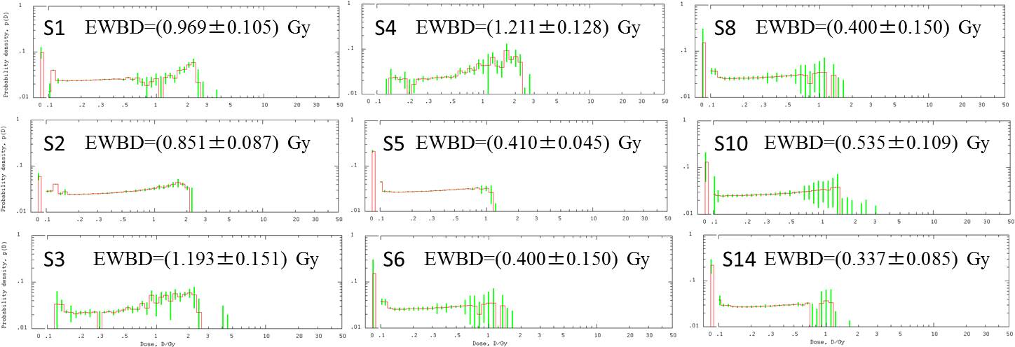
| Commentary: Dose distribution profiles as assessed by unfolding dicentric distribution (EWBD: equivalent whole-body dose) |